Solutions
Focused Ion Beam
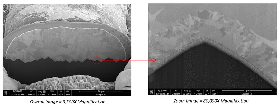
Focused Ion Beam (FIB) techniques are used in a variety of applications. In terms of failure analysis, FIB techniques are commonly used in high magnification microscopy, die surface milling or cross-sectioning, and evenmaterial deposition.
A FIB system works very similarly to a scanning electron microscope, except that it uses a finely focused beam of gallium (Ga+) ions instead of the latter’s use of electrons. This focused primary beam of gallium ions is rastered on the surface of the material to be analyzed. As it hits the surface, a small amount of material is sputtered, or dislodged, from the surface.
The dislodged material may be in the form of secondary ions,atoms, and secondary electrons. These ions, atoms, and electrons are then collected and analyzed as signals to form an image on a screen as the primary beams scans the surface. This image forming capability allows high magnification microscopy.
The higher the primary beam current, the more material is sputtered from the surface. If only high-mag microscopy is intended, only a low-beam operation must be employed. High-beam operation is used to sputter or remove material from the surface, such as during high-precision milling or cross-sectioning of an area on the die.


